02 – A VOLCANIC ALKALI-RICH BLACK PEBBLE (BOMB) FROM SÃO MIGUEL COASTLINE, AZORES (PORTUGAL)
Ano 13 (2026) – Número 1 Artigos

https://doi.org/10.31419/ISSN.2594-942X.v132026i1a2MLC
Marcondes Lima da Costa1
Susana Horta2
1Geosciences Institute, Federal University of Pará, Belém, Brazil, marcondeslc@gmail.com
2Azores Gems and Minerals, São Miguel, Azores, Portugal, azoresgemsandminerals@gmail.com; susana.horta@live.com.pt
ABSTRACT
A black pebble collected on the shore of São Miguel in the Azores, in a tabular shape, has an obsidian coating responsible for its color, concealing an interior body of light color and microcrystalline nature and volcanic amorphous glass, showing a hyalopilitic texture, in which there are crystallites and microlites of alkali-feldspars and possibly a glassy matrix of the same composition. Consequently, it is dominated by high silica, Al2O3, and alkalis (K2O and Na2O), as well as iron content. The texture, dominated by alkali-feldspars and SiO2 minerals, suggests devitrified volcanic glass of possible trachytic nature. The obsidian coating of the same composition as the interior, but microcrystalline with amorphous portions, suggests that this is a possible trachytic volcanic bomb that may have reached the ocean floor or the seashore over brachiopod bodies.
INTRODUCTION
The coast of the island of São Miguel, the largest of the nine islands of the Azores archipelago, formed by submarine volcanic activity, located in the macro-domain of the North Meso-Atlantic Ridge, receives, accumulates, and removes rocky and mineral debris resulting from the physical degradation by waves on the slopes of this island and even removed from the other islands or, less commonly, from other sources, mainly anthropogenic waste. This debris includes material ranging from medium to coarse sand, pebbles, and boulders, which are generally black, dense, and even magnetic. However, depending on the location, it may include whitish and light debris, such as pumice and volcanic slag, or yellowish to greenish, dense debris. Most are sub-rounded to angular, with a strong shine, while others are matte, sub-rounded, black to gray with a dark cortex. Among these countless pebbles, one stood out, black, with an intense to matte shine, of tabular, somewhat rounded appearance, with a slight depression with possible marks suggestive of the external surface of a brachiopod. This pebble specimen was subjected to mineralogical and chemical analyses to identify its composition and relationship with the local geology rocks, being predominantly formed by basaltic volcanic material, with dacitic to trachytic peralkaline manifestations (Ridolfi et al., 2003; Zanon, 2015; Nazzareni et al., 2019).
MATERIALS AND METHODS
The black pebble, a term that will be used throughout this article, was subjected to macroscopic and mesoscopic observations, density determination using a hydrostatic balance, analyses by transmitted light polarization optical microscopy on thin section, handheld digital microscope, chemical analyses with handheld X-ray fluorescence (XRF), scanning electron microscopy (SEM) with energy-dispersive spectroscopy (EDS). All these analyses were carried out at the LAMIGA laboratories of IG/PPGG/UFPA, in Belém, Pará. A Leica DM 2700 P optical microscope with software and digital camera, a Bruker Titan 800 XRF, a Hitachi TM 3000 SEM, and an Oxford SwiftED 3000 EDS were used. The SEM, EDS, and XRF analyses were performed directly on the natural surfaces of the black pebble and on a sawn flat surface.
RESULTS AND DISCUSSIONS
Macro to Mesoscopic Characteristics
As the name itself suggests, the pebble is intensely black in color, with a vitreous luster, a flattened tabular shape, and a slightly quadrangular outline, about 1.8 x 1.8 cm, and 3 to 5 mm thick, rounded corners, and a semi-flat surface, but with some millimeter-sized depressions (Figure 1). In the most prominent, grooves emerge that resemble features of the brachiopod shell surface (likely commissures, lines where valves meet) about 5 mm long (Figures 1 and 2). The surface ranges from smooth to randomly grooved (Figure 2). However, the black color is due to a black coating of <1 mm up to 4 mm thick, solid, conchoidal microfracture, reminiscent of obsidian (Figure 3). The interior is light-colored and microcrystalline. The density is around 2.47 and the hardness is about 6, compatible with volcanic glass. It is weakly magnetic. Overall, it is almost solid black, but locally a white mineral aggregate is identified as filling a cavity (Figures 1 and 2), an amygdaloidal type. These mesoscopic aspects, low density, and considering the volcanic nature of the insular environment, the black pebble is compatible with volcanic material, in the direction of glass, in part.
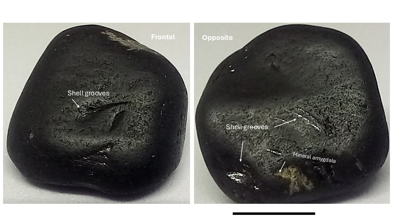
Figure 1 – The two main surface faces of black pebble from São Miguel, Azores, Portugal.
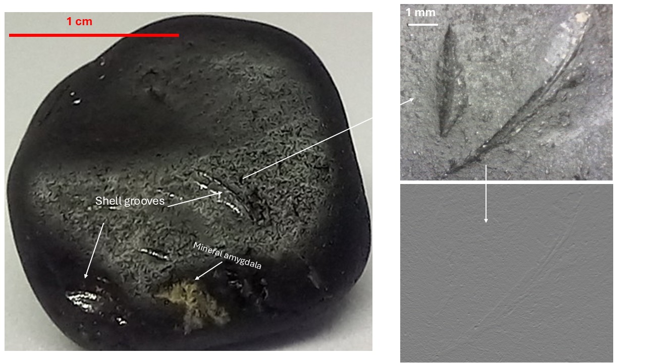
Figure 2 – Surface details which detach massive material, like a glass, slightly polished to random grooves and arched linear converging grooves suggestive of bivalve impressions (dashed rectangles). On the frontal edge, a small amygdala with whitish acicular mineral stands out.
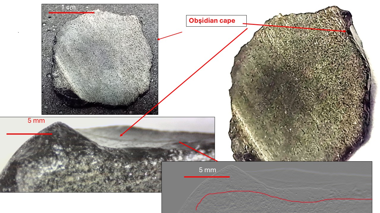
Figure 3 – Details of the obsidian cape (cortex, coating) of the black pebble.
Petrographic Characteristics under Optical Microscopy
Under transmitted light optical microscopy in thin section, the black pebble clearly shows the amorphous glassy texture of the black outer layer, obsidian-type, and the microcrystalline interior body with volcanic glass texture (Figure 4). Obsidian is already known in São Miguel and was partly described by Costa et al. (2023) but not as a coating of “pebbles.” The groundmass presents a hyalopilitic texture, in which the feldspar microlite crystals may be parallel, reticulated, radial or dendritic, spherulitic or unevenly organized (Figures 4, 5, 6). Micrometric feldspar crystals appear isolated in the glassy groundmass and glass shards can also be observed (figure 7). Iron oxide stains may be dispersed or present in the core of the reticulated or radial microlithic aggregates (Figure 5). They are usually on the order of tens of microns but can also appear as inclusions within dispersed feldspar crystals. Aggregates of radial or reticulated crystallites appear in micrometric portions of obsidian, sometimes with a core of Fe oxide stain, locally resembling spherulites (Figure 5). Perlite-type features can be locally delineated (Figures 5 and 8).
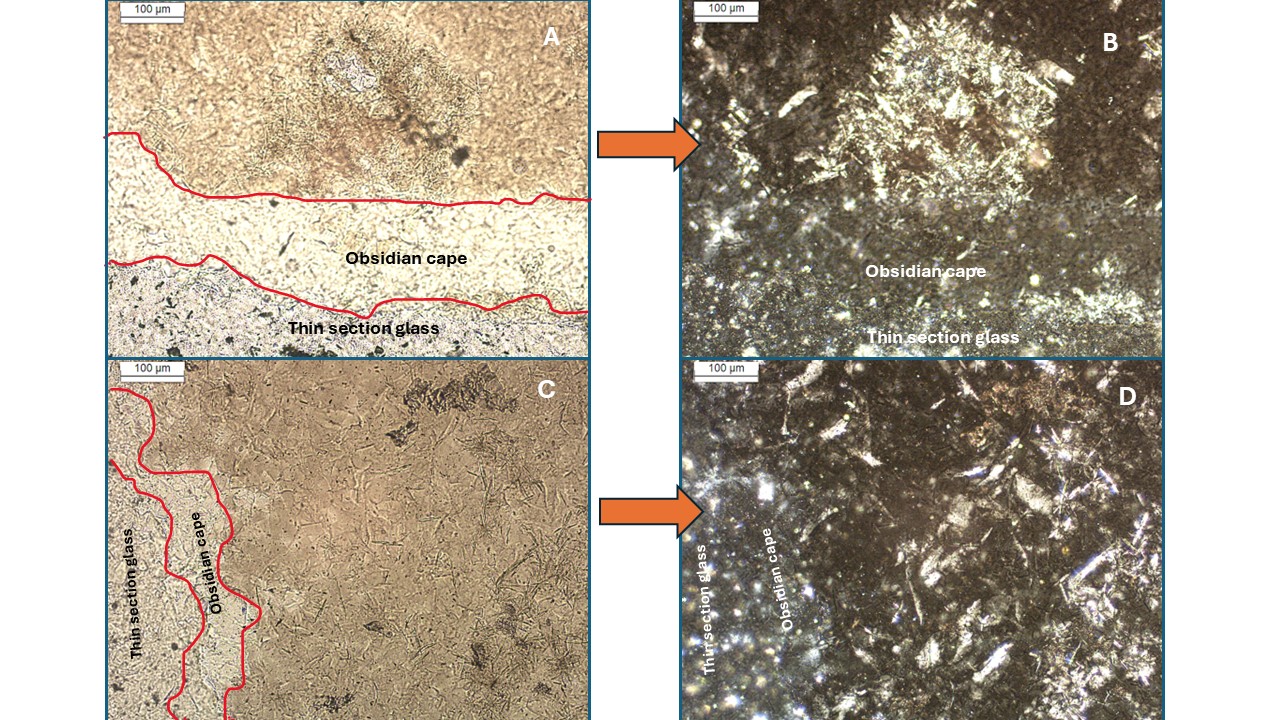
Figure 4 – A) the two areas of the black pebble under the optical microscope in natural light: obsidian cape and inner body showing a spherulite with microlites in a groundmass of alkali-feldspar crystallites and glassy material; B) the same under crossed Nicols highlighting the reticulated microlites; C) another view of the obsidian cape and inner body showing the feldspar microlites and iron oxide stains and under crossed Nicols (D).
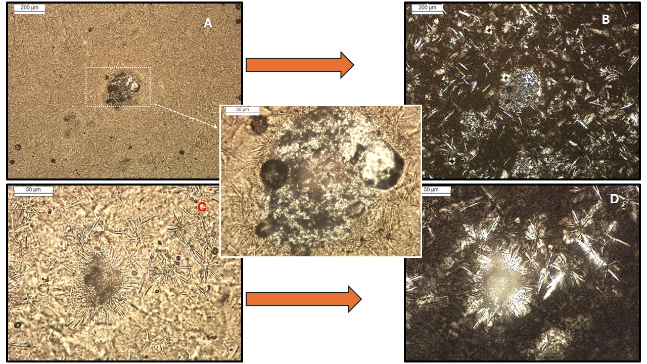
Figure 5 – A) a hyalopilitic texture detaching a spherulite made of crystallite and glass; B) details under crossed Nicols detaching the reticulated pattern of the feldspar crystallites; C) spherulite with radial crystallites, natural light; D) by crossed Nicols highlighting the feldspar crystallites and glass surrounding.
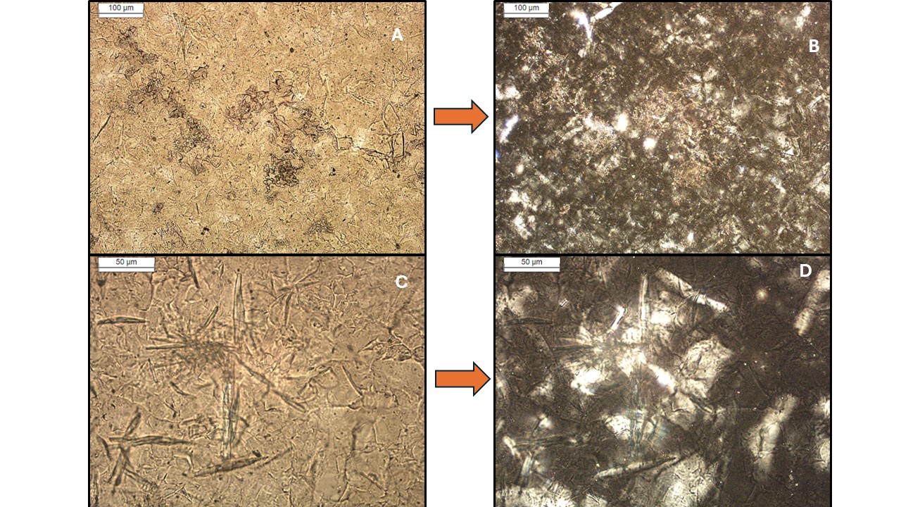
Figure 6 – A) a hyalopilitic texture detaching microlithic stains and spherulites, which stand out very well under crossed Nicols; (C) detail of sub radially forming crystallites, which becomes very evident under crossed Nicols (D).
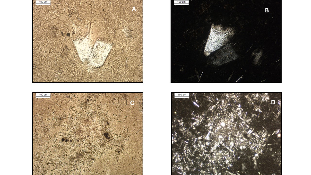
Figure 7 – A) a rare well-formed micro phenocryst of feldspar in hyalopilitic texture, natural light; B) crossed Nicols; C) microaggregates of spherulites with microlites and Fe oxide spots in the core; D) under crossed Nicols showing devitrified glass shards into alkali-feldspars.
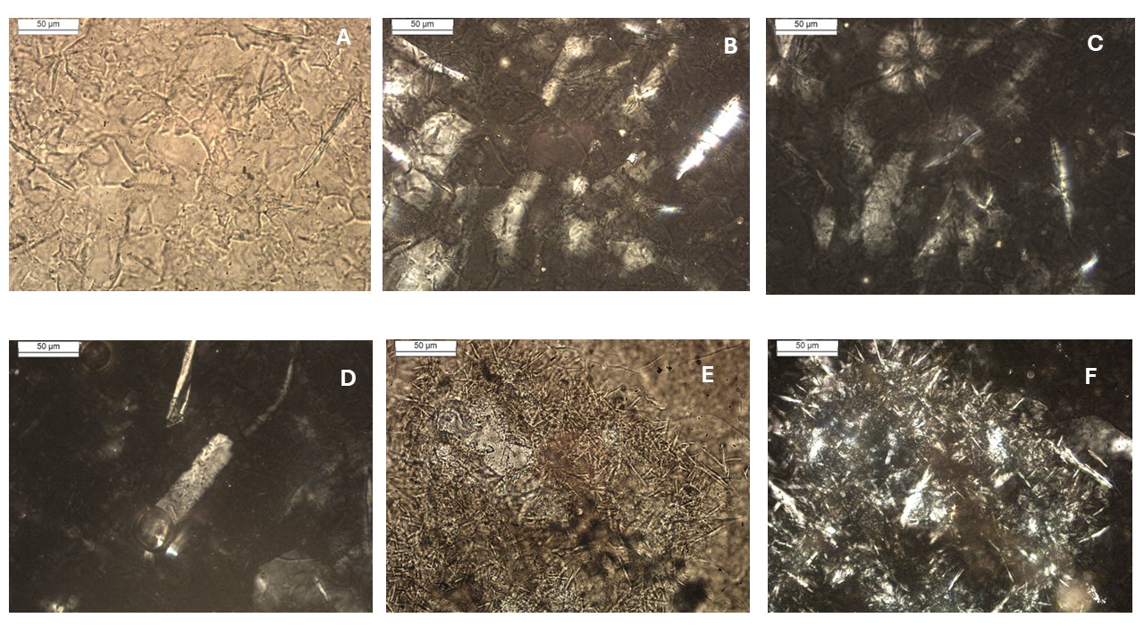
Figure 8 – A) Detail of the amorphous to microcrystalline groundmass and pearlitic mass (irregular to pseudohexagonal outlines with microlites, natural light, which in crossed Nicols highlight tabular shapes of alkali-feldspars and perhaps opal and/or quartz (B), which can converge into radial spherulite of the same mineralogy (C), then a tabular alkali-feldspar crystal in a glass domain (D); E) a typical spherulite with sub radial microlites and perlitic aspect and vitreous spots of Fe oxides; F) image E in crossed Nicols with devitrified glass shards.
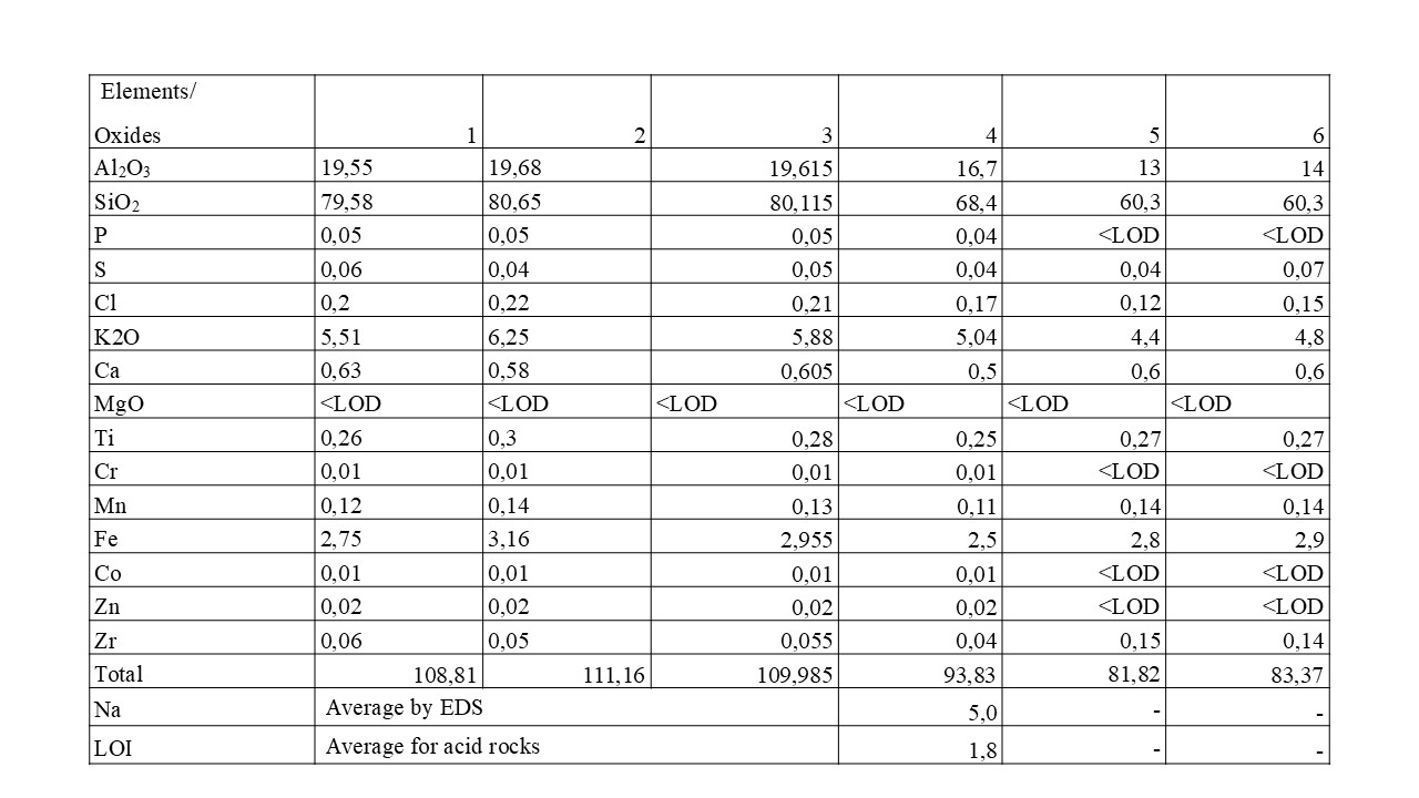
Semi-quantitative Chemical Composition according to XRF
The corrected results of the semi-quantitative analyses on the two main faces show similar results, with a dominance of SiO2 (~68.4% by weight), Al2O3 (~16.7%), which is comparatively high for igneous rocks, as well as notable amounts of K2O (~5%), Fe (~2.5%), followed by Ca (~0.5), Ti (0.25%) and Cl (0.2%), with MgO < 0.5 (LOD) (Table 1). These results indicate that it is a rock of intermediate to acidic nature, rich in silica, alumina, and alkalis (K, Na). The SiO2 contents suggest the presence of quartz and/or opal as constituent minerals. The alkalis, aluminum, and silica suggest the presence of alkali feldspars and quartz/opal, possibly. The black color of the pebble, the obsidian cape, may reflect the Fe content as opaque minerals, such as oxides. The low contents of Ca, Mg, Ti, V, Cr, Mn, Ni exclude rocks of mafic nature.
Table 1 – Semiquantitative chemical composition after handheld XRF. Sodium was not detected due to method limitations, but as will be shown by SEM/EDS, it is present, probably at potassium levels. These values were corrected to 100%, considering the Na content (5%) and LOI (1.8%). (1. On the uneven black glass surface, front face; 2. On the uneven black glass surface, opposite face; 3. Average; 4. Corrected to 100%. 5. On the flat sawn surface, microcrystalline; 6. On the flat sawn surface, microcrystalline). <LOD: below the detection limit of handheld XRF; – not analyzed.
Microfabric and Mineral-Chemistry after SEM/EDS
On a micrometric scale under scanning electron microscopy, the black pebble shows features of elongated tabular to irregular crystals, ranging from 20 to 50 µm, with partly jagged edges or corrosion features, in mass formed by aggregates on the order of 5 to 10 µm, irregular to cylindrical (Figure 9). Numerous chemical analyses suggest (K, Na)-feldspars, orthoclase/sanidine type. Irregular portions in dark gray shades, ranging from 5 to 50 µm, massive, suggest amorphous material, glass-like. Its chemical composition (Fig. 9 analyses 1 to 5; Fig. 10 analyses 2 to 4; Fig. 11 analyses 1 to 4; Fig. 12 analyses 1 to 3) corresponds to amorphous orthoclase/sanidine, with compounds of Ca, Cl, S, and Fe. The high levels of Na and Cl observed in the dark gray spots, corresponding to the amorphous material, consistent in all of them, suggest the presence of Na chloride, halite type, and of S and Ca, gypsum. Crystals with fractures and conchoidal surfaces (Figure 10) are also observed, with circular to irregular outlines; frequent ovoid aggregates, tabular fragments, glass (dark gray), and irregular and cylindrical microaggregates (Figure 10), in addition to subtle features that suggest possible perlite (Figure 11). Consistent Fe content values ranging from 2 to 14 in the orthoclase/sanidine domain indicate the presence of Fe oxide inclusions in micro- and nanometer sizes. The excess oxygen values, along with Si, mainly in the spots, were related to the presence of opal, or even tridymite and/or cristobalite. This mineralogy is present in volcanic glass, partially to fully devitrified (Zarins, 2025). Micros can be recognized in these SEM images and analyzed locally. It is worth noting the presence of Ca, S, Cl, and rarely C, in the glass domains (dark gray spots). The dominance of alkali-feldspars in the groundmass and the presence of rare micro phenocrysts point to a trachytic composition domain, associated with explosive material (Zanon, 2015). Microcrystalline fragment- or ovoid-type features also stand out in the groundmass and present the same chemical-mineralogical composition (Figures 12 and 13).
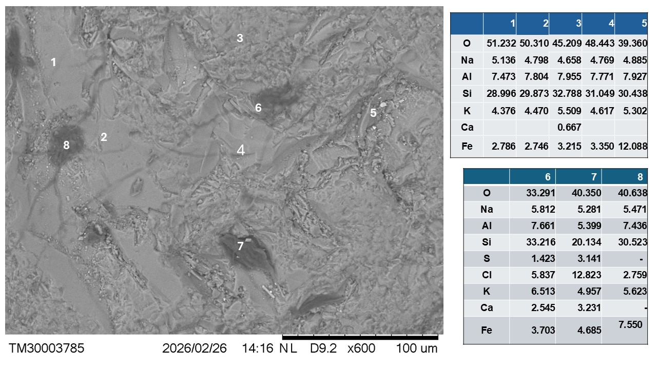
Figure 9 – Electron photomicrography (SEM) and semiquantitative chemical analyses (EDS) of the microgranular to glassy groundmass, showing the dominance of crystalline orthoclase/sanidine (light gray) and amorphous (dark gray matter = glass) and perlite-type circular features.
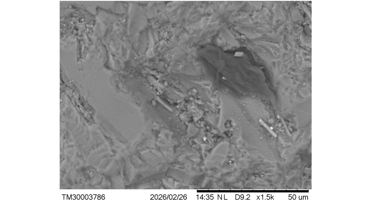
Figure 10 – A possible phenocryst with a corroded rim, containing interstitial microlites and cylindrical features, and typically glassy material with a chemical composition corresponding to the orthoclase/sanidine domain; micrometric light gray spots are composed of iron oxides.
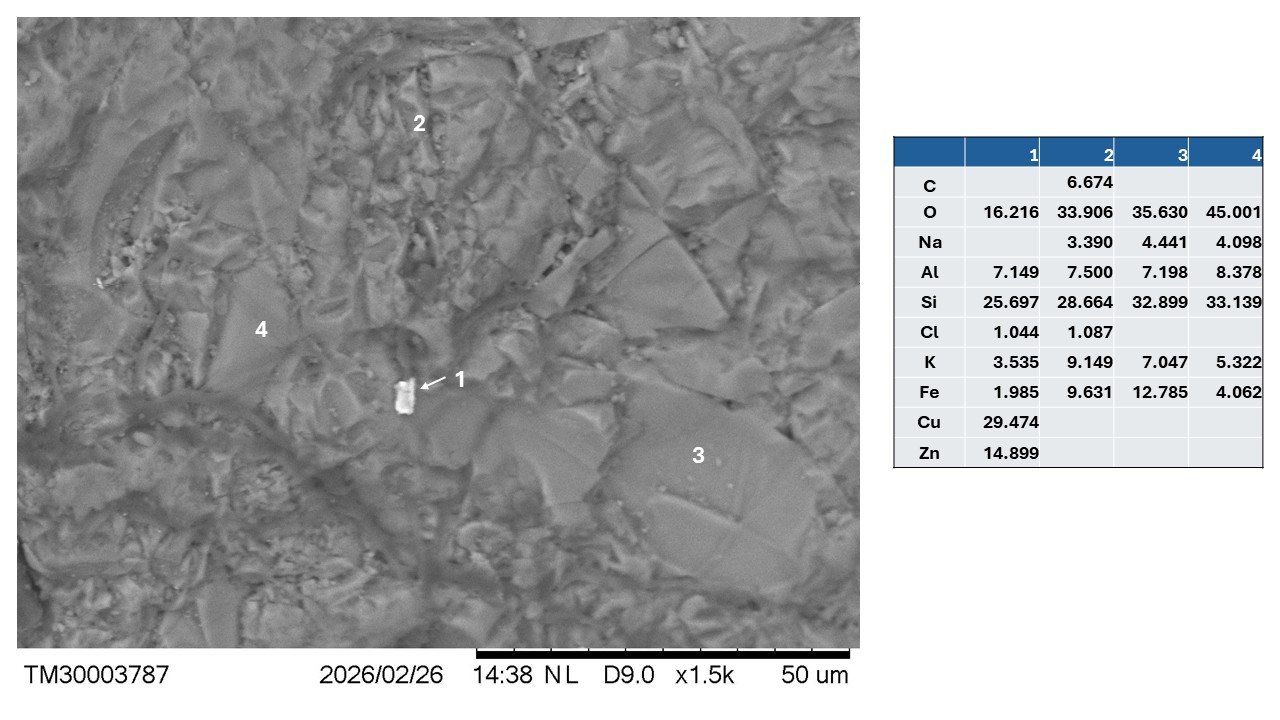
Figure 11 – Groundmass with small crystals exhibiting conchoidal fracture with higher potassium content, similarly to iron (analyses 2 to 4) and the rare presence of Cu-Zn alloy (analysis 1).
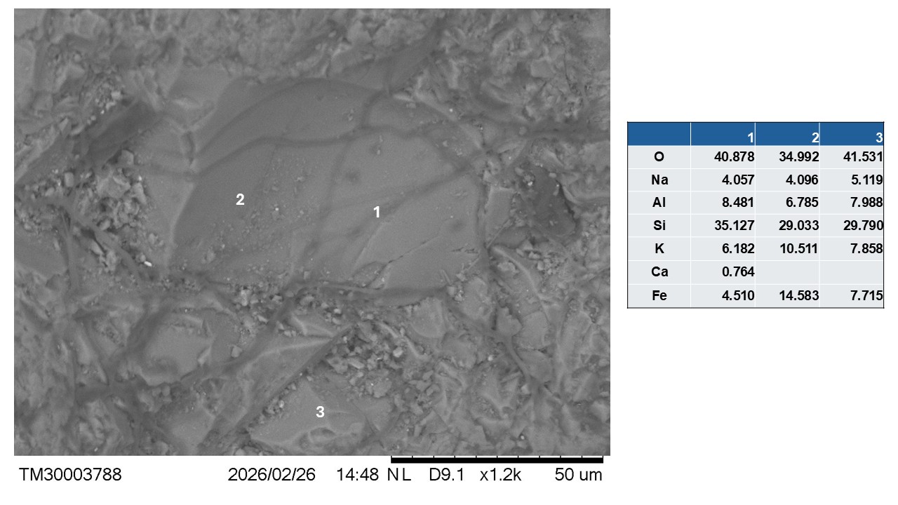
Figure 12 – Crystal or suboval fragment, fissured and with conchoidal fracture in a microcrystalline domain with glassy zones, but with a chemical composition in the orthoclase or orthoclase/sanidine range with high and variable iron content.
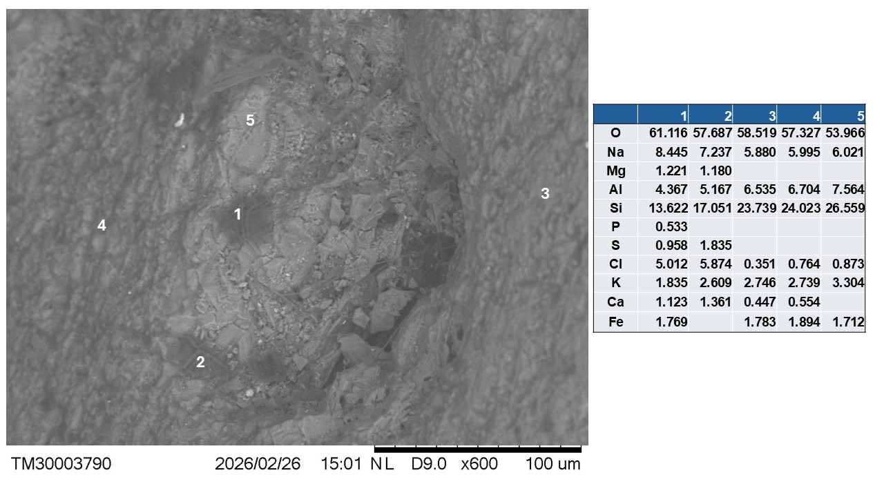
Figure 13 – Ovoid microcrystalline fragment or aggregate (analysis 5: albite and orthoclase + NaCl, Fe oxides) with glassy portions (analysis 1) shaded and in sub microcrystalline groundmass (analyses 3 and 4, like 5) also containing glassy spots (analysis 2 comparable with 1).
Although rare, particles with very irregular shapes and slightly aligned, ranging from < 1 µm to about 15 µm (Figures 14 and 15, analyses 1 and 2) of alloy (Au-Cu-Ag) were locally detected in the groundmass of orthoclase + opal (excess O and Si).
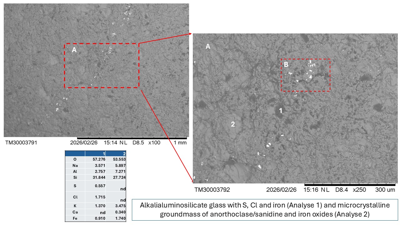
Figure 14 – Linear aggregates of irregular and submicrometric and micrometric particles of (Au-Cu-Ag) alloy (A) in the domain of orthoclase + opal (A, 2) and glass (A, 1). The B rectangle is showing in the next figure 15 with Au-Cu-Ag alloy SEM-EDS analyses.
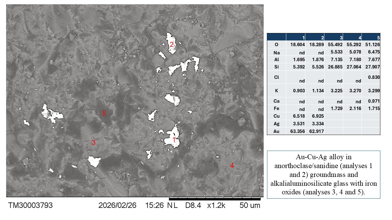
Figure 15 – Detail of the disform and submicrometric to micrometric particles of (Au- Cu-Ag) alloy with SEM-EDS analyses.
Aggregates of micrometric to submicrometric titanomagnetite particles, disordered, also appear rarely in droplet or tabular form, encompassing material with composition corresponding to orthoclase/sanidine, glass, and opal, and possibly apatite (Figures 16 and 17), always in the groundmass of orthoclase/sanidine + Fe oxides. The chemical composition of these titanomagnetite aggregates varies very little and seems to incorporate Mn and Mg, perhaps Al, with partial equivalence to the titanomagnetites described by Zhu et al. (2023) in the Panzhihua ore, which, however, are vanadium-bearing, associated with ilmenite and magnetite and included in olivines and pyroxenes, being more characteristic of mafic and ultramafic rocks. However, titanomagnetites can be also found in volcanic rocks ranging from basaltic to trachytic, dacitic, and andesitic (Yousef et al., 2024), and the Azores archipelago involves volcanic rocks from basaltic to peralkaline (Ridolfi et al., 2003; Zanon, 2015; Nazzareni et al., 2019).
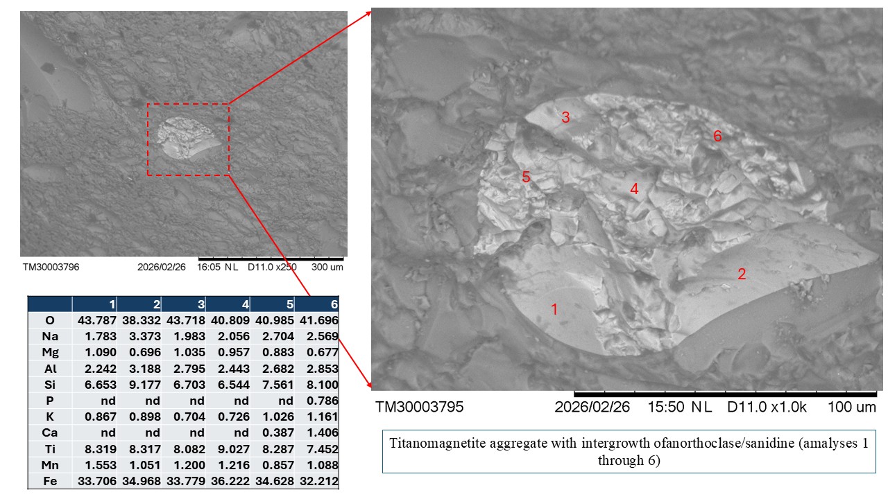
Figure 16 – Aggregated in droplet form (100 µm large) of submicrometric and micrometric titanomagnetite particles intergrown with material of orthoclase/sanidine composition and Fe oxides in the alveolar groundmass of this composition.
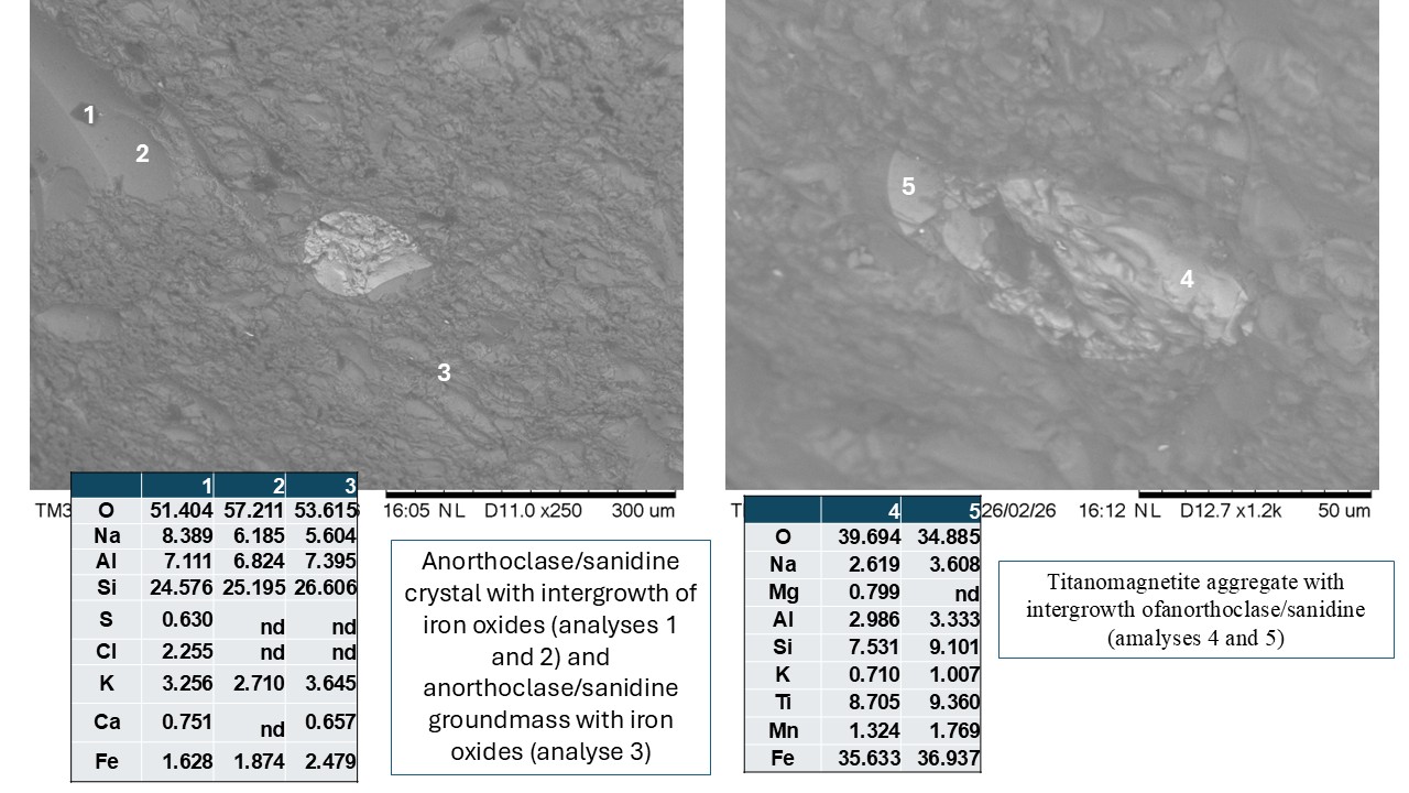
Figure 17 – Probable reabsorbed alkali-feldspar crystal with glassy spots with titanomagnetite aggregates in droplet and tabular form (analyses 4 and 5) in the alveolar groundmass (analysis 3) (figure on the left and right, respectively).
CONCLUSIONS
The black pebble investigated here is tabular, low-density, and formed by a thin black glassy cape or cortex equivalent to obsidian, which surrounds microcrystalline to amorphous material of a lighter color. Thus, the external black color is given by the obsidian cape. The chemical composition of the cape corresponds to material of alkali-feldspar nature with iron oxides and opaline. The chemical composition of the enclosed material is like that of the cape, but predominantly microcrystalline, with many glassy-amorphous micro spots, showing typical volcanic textures, hyalopilitic, including devitrified volcanic glass, such as devitrified spherulites with feldspar microlites and possibly quartz and/or quartzine, opals, radial textures, conchoidal fractures, and perlitic textures. The contents of SiO2, Al2O3, and Na2O+K2O correspond to the composition of alkaline rocks, like trachyte. Iron oxide particles, such as spots and in micrometer to sub micrometer sizes, are frequent, especially the sub micrometer ones, perhaps even nanometric, which are intergrown with alkali-feldspar aggregates and spherulites. They also contain rare inclusions of (Au-Cu-Ag) and (Cu-Zn) alloys, which form irregular micrometer-sized particles as well.
The black pebble investigated probably retains much of its original external morphology and dimensions; that is, although found on the shoreline, it would have been little polished (reworked) by marine physical activities, since it still preserves its structure that suggests a possible trachytic volcanic bomb. This is further reinforced by the probable impression of micro brachiopod shell commissures. It can be assumed that bombs like the black pebble, still in a hot magma-pasty form, might have reached these benthonic organisms located on the coastal seabed or even on the beach line; this could be an explanation. Many pebbles from the São Miguel shoreline with these much more striking impressions were collected by the second author, S. Horta, and are currently under study and cited in her chronicle published in this BOMGEAM issue (Horta, 2026): SOLITE: A “PEDRA DA RESILIÊNCIA” O Enigma do Carbono e a Identidade das Ilhas de Bruma.
Acknowledgments
To CNPQ for granting financial support through grant number 304.967/2022-0 and to the lamination (thin section) sector of IG/UFPA. All analyses were carried out by the first author in the LAMIGA laboratories of PPGG/UFPA and the field activities by the second author with support from Azores Gems and Minerals.
REFERENCES
Costa, M.L. Horta, S., Santos, P.H.S., Valente, G.J.S., Pinto, D. C. 2023. An obsidian from Lagoa Sete Cidades Lagoon on São Miguel island, Azores, Portugal. BOMGEAM, 10 (3); DOI 10.31419/ISSN.2594-942X.v102023i3a5MLC.
Horta, S. 2026. SOLITE: A “PEDRA DA RESILIÊNCIA” O Enigma do Carbono e a Identidade das Ilhas de Bruma. BOMGEAM, 13 (1). Preprint.
Nazzareni, S., Nestola, F., Zanon, V., Bindi, L., Scricciolo, E., Petrelli, M., Zanatta, M., Mariotto, G., Giuli, G. 2019. Discovery of moissanite in a peralkaline syenite from the Azores Islands. Lithos, 324–325: 68-73.
Ridolfi, F., Renzulli, A., Santi, P., Upton, B.G.J., 2003. Evolutionary stages of crystallization of weakly perlakaline syenite: evidence from ejecta in the plinian deposits of Agua de Pau volcano (São Miguel Azores Islands). Mineralogical Magazine, 67(4), 749-767.
Yousef, F., Lentz, D.R., and Bark, G. 2024. Titanomagnetite Rims on Hornblende Phenocrysts in Intermediate Subvolcanic Intrusions, Torud–Ahmad Abad Magmatic Belt, Iran: Examination of Devolatilization and Oxidation Processes. Lithosphere Volume 2025, Number 1, Article ID lithosphere_2024_227, 13 pages https://doi.org/10.2113/2025/lithosphere_2024_227.
Zanon, V. 2015. The magmatic suite of the Azores. In: Gaspar, J. L., Guest, J. E., Duncan, A. M., Barriga, F. J. A. S. & Chester, D. K. (eds) Volcanic Geology of Sa ̃o Miguel Island (Azores Archipe-lago). Geological Society, London, Memoirs, 44, 51–64, http://doi.org/10.1144/M44.5.
Zarins, A. 2025 The geology of agate deposits. Springer, Cham, Switzerland, 158p.
Zhu, F.; Ma, Z.; Gao, G.; Qiu, K.; Peng,W. Process Mineralogy of Vanadium Titanomagnetite Ore in Panzhihua, China. Separations 2023, 10, 147. https://doi.org/10.3390/separations10030147.