04 – MINERALOGICAL AND TEXTURAL CHARACTERISTICS OF GRAÇA CHALCEDONY IN PALEOZOIC SEDIMENTARY ROCKS OF PARNAÍBA BASIN (STATE OF MARANHÃO, BRAZIL)
Ano 06 (2019) – Número 02 Artigos
![]() 10.31419/ISSN.2594-942X.v62019i2a4THBLS
10.31419/ISSN.2594-942X.v62019i2a4THBLS
Características mineralógicas e texturais das calcedônias Graça em rochas sedimentares paleozoicas da bacia do Parnaíba (Maranhão, Brasil)
Tauan Henrique Bittencourt Lima da Silva1,
Rosemery da Silva Nascimento2,
Marcondes Lima da Costa2,
Érico Rodrigues Gomes3
1 Under graduating geology student, Federal University of Pará, Belém, Pará, Brazil,thj272@hotmail.com
2 Geoscience Institute, Federal University of Pará, Belém, Pará, Brazil, marcondeslc@gmail.com, rsn@ufpa.br
3 Federal Institute of Science and Technology, Teresina, Piauí, Brazil, ericorg66@gmail.com
ABSTRACT
Chalcedony is a fibrous cryptocrystalline variety of quartz normally found in volcanic, hydrothermal and sedimentary environments. In the Parnaiba sedimentary basin in northeastern Brazil, Paleozoic sedimentary rocks of Pedra de Fogo Formation carry several occurrences of such materials, which are clearly related to deeply silicified mudstones, siltstones and even sandstones of this geological unit. A very interesting occurrence was found in São João dos Patos region, southeast of Maranhão, at Graça farm. These chalcedonies show four most characteristic patterns: bluish white chalcedony, dendritic chalcedony, banded chalcedony, and merged chalcedony. The Graça chalcedonies are translucid and do not feature extravagant colors, and is filled with dendrites and relicts, even though those properties limit their use as jewels, they show potential on simple cuts, such as cabochon.
Keywords: Manganese Dendrites; Hollandite; Quartz; Cabochon; Agate.
RESUMO
Calcedônia é uma variedade fibrosa de quartzo criptocristalino, formada partir de processos vulcânicos, hidrotermais e sedimentar-diagenéticos. Nas rochas sedimentares da Formação Pedra de Fogo do paleozoico da Bacia do Parnaíba ocorrem calcedônias em terrenos do município de São João dos Patos, sudeste do estado do Maranhão. Essas calcedônias segundo seus aspectos texturais ser classificadas como: calcedônia branca azulada; calcedônia dendrítica; calcedônia bandada e calcedônia mesclada. Sob o microscópio ótico foi possível identificar o hábito fibroso e texturas características desse mineral. As calcedônias da Graça são translúcidas e não apresentam cores extravagantes, além de serem permeada por dendritos e relictos dos arenitos originais, que são atributos limitantes seu uso gemológico. Porém se permitem a lapidação simples, como o cabochão.
Palavras chave: Dendritos de manganês; hollandita; quartzo; cabochão; ágata.
INTRODUCTION
Chalcedony is a general term that refers to the microcrystalline fibrous α-quartz. Chalcedony commonly occur associated to volcanic rocks as products of post volcanism alteration in hydrothermal veins, fractures in crystalline rocks or pores and concretionson sedimentary rocks, as a result of low temperature (<100ºC) and SiO2– rich fluid percolation (Moxon 2002, 2009). Fluctuation of temperature and chemistry changes during chalcedony formation will change the mineral itself, it is not unusual the formation of bands of alternating composition. Banded chalcedony is known as agate, and this feature is caused by various occurrences: Fe oxide spots, apparent pauses in the crystallization process and textural changes. Less common is the planar banding that is presumed to have been gravity-controlled (Moxon & Reed 2006).
The first reported use of chalcedony comes from Mesopotamia, where tools and adornment were found, the most famous is an axe head, dated 3000 B.C. and was also mentioned in the Bible (Revelation 21:19, 21:20) (Vilasbôas et al. 2017). It is known that chalcedony has been used by humankind since the dawn of society, as hunting weapons and other tools, until today, as lab equipment, jewelry, ornaments and others.
In Brazil, the most famous chalcedonies and agates deposits are those of Soledade, Salto do Jacuí, Iraí, Ametista do Sul and Frederico Westphalen, all of them in Rio Grande do Sul. There are also small deposits from Minas Gerais, Bahia, Goiás and recently the Fazenda Calçadinha deposit, also occurring on Nazareth do Piauí (Costa & Silva 2016).
A new chalcedony occurrence was found by the Geologist Érico Gomes and the Prof. Dr. Marcondes Lima da Costa in the farm of Mrs. Graça, thus, the name, (São João dos Passos locality, São João dos Patos municipality, Maranhão) (Figure 1).

Figure 1 – Location map of São João dos Patos municipality, including neighboring locations, the Boa Esperança Dam, and its geology.
The region around the Graça chalcedony is developed on fine white friable sandstones and siltstones of Pedra de Fogo Formation from the Parnaíba Basin, exhibiting concretions of semi-translucent, cohesive, varicolored chalcedony, besides fossil wood trunks made of chalcedony too.
The Pedra de Fogo Formation is a marine regression, with prevalent continental sedimentation in a period of pronounced aridity (Vaz et al. 2007, Santos & Carvalho 2009). There are many references about the occurrences of chalcedony in the region, except for that on fossil trunks. Araújo and Costa published a book about the Fossil forest at Tocantins, widely referencing chalcedony (Martins, 2010). The paper Costa & Silva wrote in 2016 determined that this Formation should be a potential environment for chalcedony occurrence, such example is the Calçadinha chalcedony.
This article described the mineralogical and textural features of Graça chalcedony and gemological potential.
MATERIALS AND METHODS
Initially a geological survey supported by GPS and Google Earth was executed by Prof. Dr. Marcondes Lima da Costa and geologist Érico Rodrigues Gomes. During field work eight samples of chalcedonies were collected, and then transported to the Cutting Laboratory of UFPA, crafted into six thin sections and analyzed at the Petrography Laboratory of the Geoscience Institute of UFPA, on AXIO Scope1 microscope up to 30X increase, in order to describe the mineralogical and textural aspects of Graça chalcedony. The images were taken by the LEICA Application Suite V 4.6 software coupled to a LEICA DM 2700 P microscope. To support the descriptions and images, the thin sections were analyzed under SEM/EDS and Microraman, while the hand samples were cut and polished, furthering gemological appreciation.
The semi-quantitative were carried out with Energy Dispersive Detector (SEM/EDS) coupled to Scanning Electron Microscope with the objective of determining the mineral inclusions as well as microstructure images of the distinct chalcedonies. The SEM model used is a LEO-1430 linked with a EDS Sirius-SD system. The thin sections were metallized with gold. The images were generated under electron beam of 90μÅ, constant acceleration voltage of 20 kV, and working distance of 15 mm.
Additional microraman analyses were carried out with equipment from the Geoscience Museum of Federal University of Pará, of the Gem Expert Brand, BWTEK model equipped with the Gem ID Identification software.
For gemological purposes, the chalcedonies were polished on the Cutting Laboratory of UFPA by use of a carborundum polishing stone; the samples that acquired the best luster were taken to São José Liberto Museum Gemological Laboratory, where they were faceted and polished into simple, plain cuts, five cabochons, shaped as three triangles, one sphere and one dome. As well as two faceted cuts (fantasy and emerald). The chalcedonies were cut by a circular saw, based on the expected stone size, and cooled by water. The first five gems were finely polished on a stoneware wheel, to acquire cabochon shape, the last two were glued to a polishing pen, and faceted according to specific angles by inserting this pen into a holed board near a polishing disc.
RESULTS AND DISCUSSIONS
The Graça chalcedony manifests itself in several colors, with different textures and minerals, these characteristics enabled the classification of four chalcedony varieties:
- White bluish chalcedony, with sparse dendrites and cohesive matrix.
- White dendritic chalcedony, with friable matrix.
- Banded vesicular chalcedony, alternating red and grey layers.
- Black and white interspersed chalcedony.
The white bluish chalcedony tends to manifest some sparse Mn dendrite. This type is made of a core of chalcedony, bordered by milky white opal that detaches the core from the heavily silicified sandstone matrix. Fe oxyhydroxides are underdeveloped and only seen in thin section, unlike Mn oxyhydroxides. Because of the distance between its dendrites, will be referred to as mosquito from here on (Figure 2A).
The dendritic chalcedony is the most common, similar to the bluish, due to their color and chalcedony core bordered by opal, where the mosquito has underdeveloped dendrites, the dendritic, as the name suggests, has well developed Mn and Fe oxyhydroxides dendrites, friable matrix, several relicts of sandstone mingled within chalcedony core, as well as holes filled with chalcedony and opal. These samples will be pointed as dendritic onwards (Figure 2B).
Banded vesicular chalcedony manifests a characteristic banding, iterating hues of red, gray and white. Scattered across its surface are many vesicles and amygdales, these filled with quartz and/or Fe oxyhydroxides, as well as spheroidal structures made of chalcedony aggregates and Fe oxyhydroxides. Owing to its banding, will be called agate from now on (Figure 2C).
Black and white chalcedony is represented by ones that mingles quartz, opal and chalcedony with the oxyhydroxides minerals of manganese and iron, either in layers or randomly, it is brittle and compact. Due to its color, will be referred to as onyx (Figure 2D).
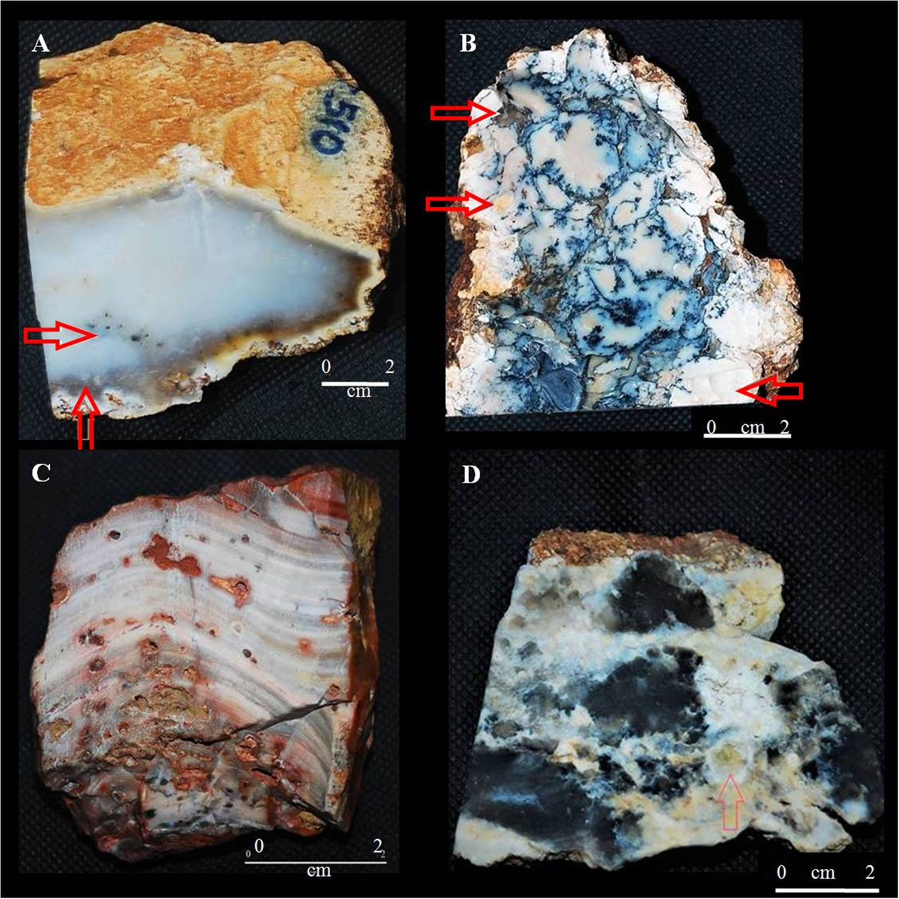
Figure 2. Images of hand samples illustrating the four recognized varieties of Graça chalcedonies. (A) Mosquito chalcedony, the white arrow points to a dendrite, the brown to the opal border. (B) Dendritic chalcedony, the red arrow points to a sandstone relict, the brown to the opal border and the white to a cavity filled with botryoidal chalcedony. (C) Agate, the image clearly shows its banding and many vesicles. (D) Onyx, the image defines the mingling between chalcedony and the dendrites, the red arrow points to a cavity filled with chalcedony and opal.
MINERALOGY
The mineralogical manifestations within Graça chalcedonies match the cryptocrystalline nature characteristic to this mineral, thus, are better analyzed with the aid of an optic microscope. The quartz, of course, is the main mineral and barite also occur, recognized only by SEM/EDS analyses and Fe and Mn oxyhydroxides.
Quartz chalcedonic
Graça chalcedony is represented by spherical, fibrous, wall-lining, cryptocrystalline aggregates and also replacing siltstones or previous quartz crystals (Figures3A, B and C) as such, they are encompassed by other mineral occurrences, like Fe and Mn oxyhydroxide dendrites, opal and quartz. The chalcedony may appear perpendicular or parallel to the eye, showing a granular or needle-like pattern (Figure3D).
The heterogeneous mineral patterns observed in Graça chalcedony allows a wide range of microtextures and structure such as massive chalcedony, banded chalcedony aggregates of cryptocrystalline quartz, chalcedony and Fe, Mn dendrites with crystalline rim, similar to moss vegetation and fine rhythmic bands in a kidney shape (Figures 3 and 4). In such features the Graça chalcedonies can be well compared to Calçadinha ones described by Costa et al (2016).
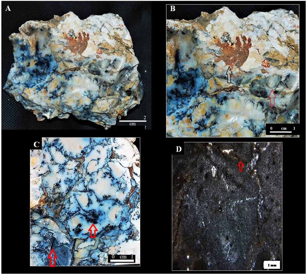
Figure 3. (A) Dendritic chalcedony with Fe, Mn dendrites and opal occurrence (B) A detail of figure A, the black arrow points to a Fe oxyhydroxide, the red arrow indicates an opal manifestation. (C) Dendritic chalcedony, the brown arrow points to hemi-spherulite aggregates of Mn oxyhydroxide, the red arrow indicates a micro druse of quartz, both manifestations within the massive chalcedony domain. (D) Mineralogical manifestations of dendrites interspersed with chalcedony and quartz, the red arrow points to a Fe dendrite and the white arrow shows a Mn oxyhydroxide.
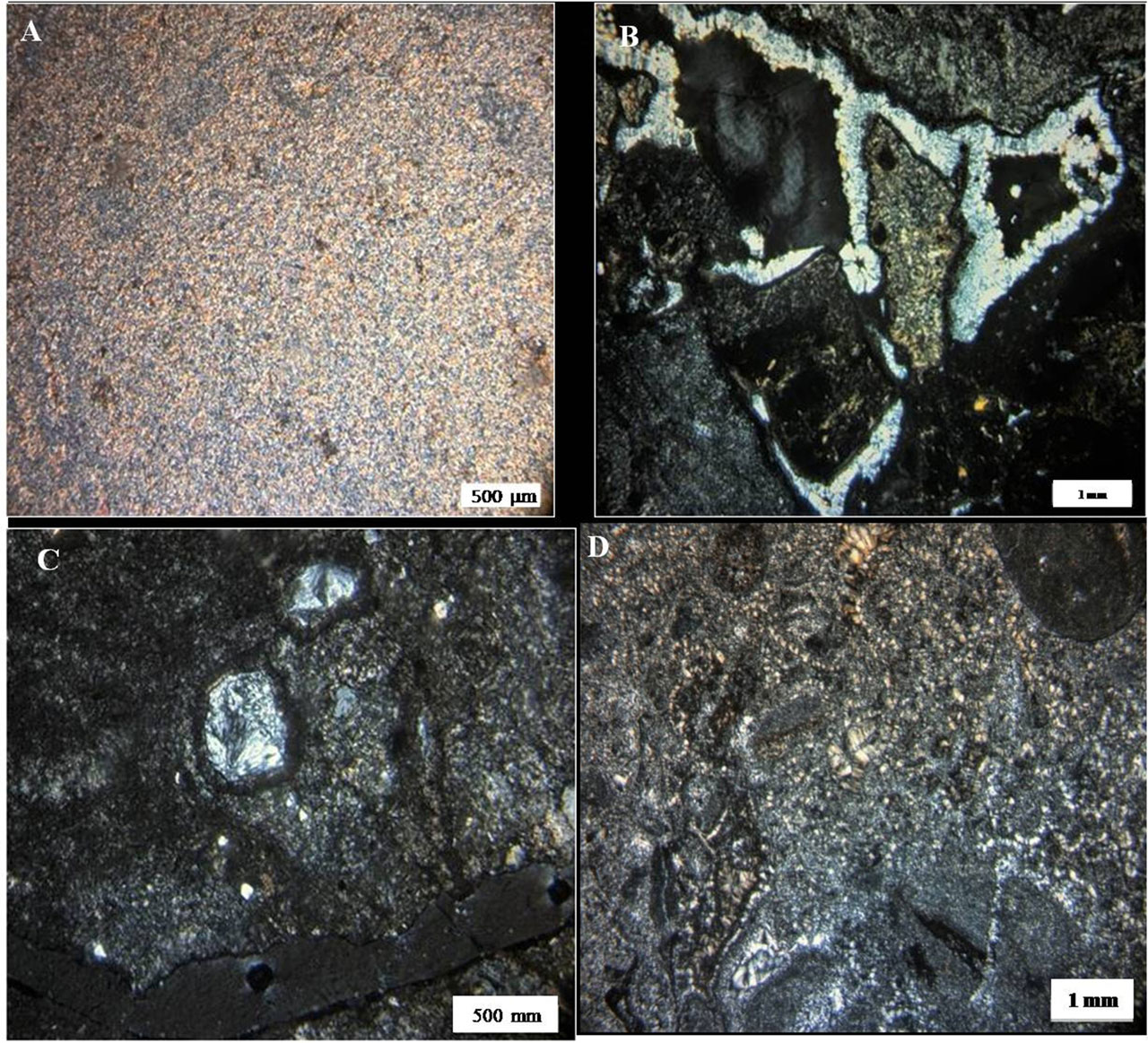
Figure 4. (A) Massive chalcedony mingled with Mn and Fe dendrites. (B) Well developed chalcedony that partially fills cavities, whilst side-by-side with underdeveloped chalcedony with Fe and Mn oxyhydroxides. (C) Chalcedony replacing a probable quartz grain, bordered by both Fe and Mn dendrites. (D)Overview of cryptocrystalline quartz matrix, the radial-needle shape is widely dominant.
Comparing SEM analyzes to microscopic, the texture and dirty appearance observed with Nichols uncrossed are caused by chalcedony micro porosity, observed in all varieties of chalcedony, these pores are about 0.1 micron in diameter, circular and scattered in the thin matrix of chalcedony.
The predominant of massive chalcedony, characterized by microcrystalline quartz aggregates, unchanged or oriented, formed under conditions of silica supersaturation at low temperatures, which led to the low crystallinity of the material (Figure5) (Morrison et al. 1995).
Subordinately occurs the colloform texture, common to chalcedony, where the crystals form in rhythmic bands, organizing themselves in reniform habit or botryoidal, when observed in plant (Figure 5A and B), this aspect is inherited from the silica that, by presenting strong surface tension, maintains the reniform aspect even after its crystallization (Morrison et al.,1995). Although this texture is common to chalcedonies, it was only observed in thin section and in SEM analyses (Figure 9).
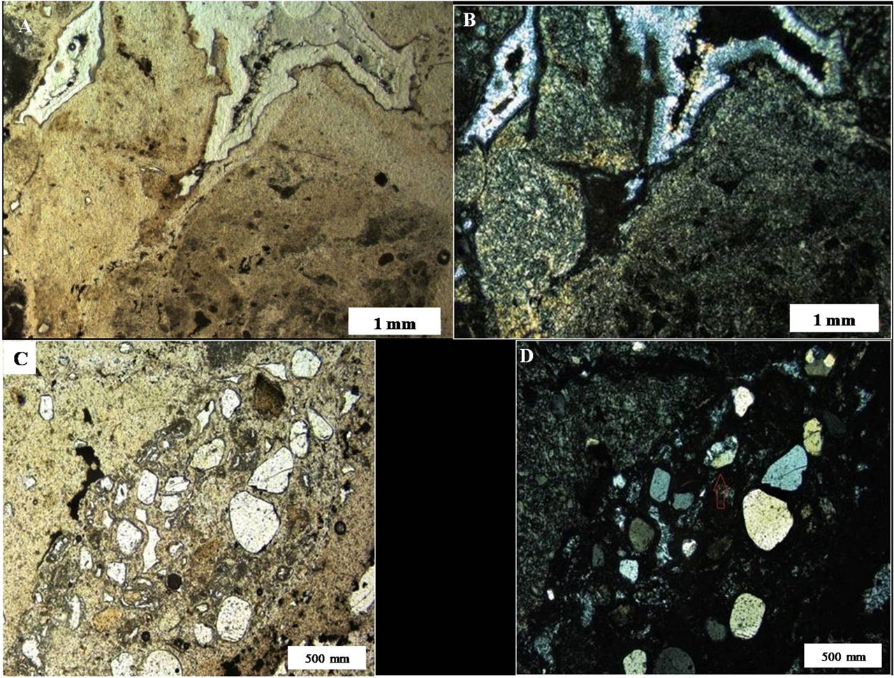
Figure 5. Colloform chalcedony with relict pattern of sandstones with Fe and Mn dendrites. (A and B). Quartz in rounded to uneven grains, some replaced partially, or completely by chalcedony.
Moss is another texture observed and limited only to the agate (Figure 8A and 8B) however. The moss texture is characterized by fine botryoidal aggregates, which resemble the vegetation namesake, with very fine quartz texture in concentric pattern at its core and thicker border, the agate variety pattern shows a spiral of chalcedony and Fe oxyhydroxides (Figure 6B and 6C) (Morrison et al. 1995).
The last texture observed is inherited, where quartz grain-sized from sandstones maintain their original granular habit, and sometimes cryptocrystalline quartz might replace partially or completely these remains (Figure 7C and 7D).
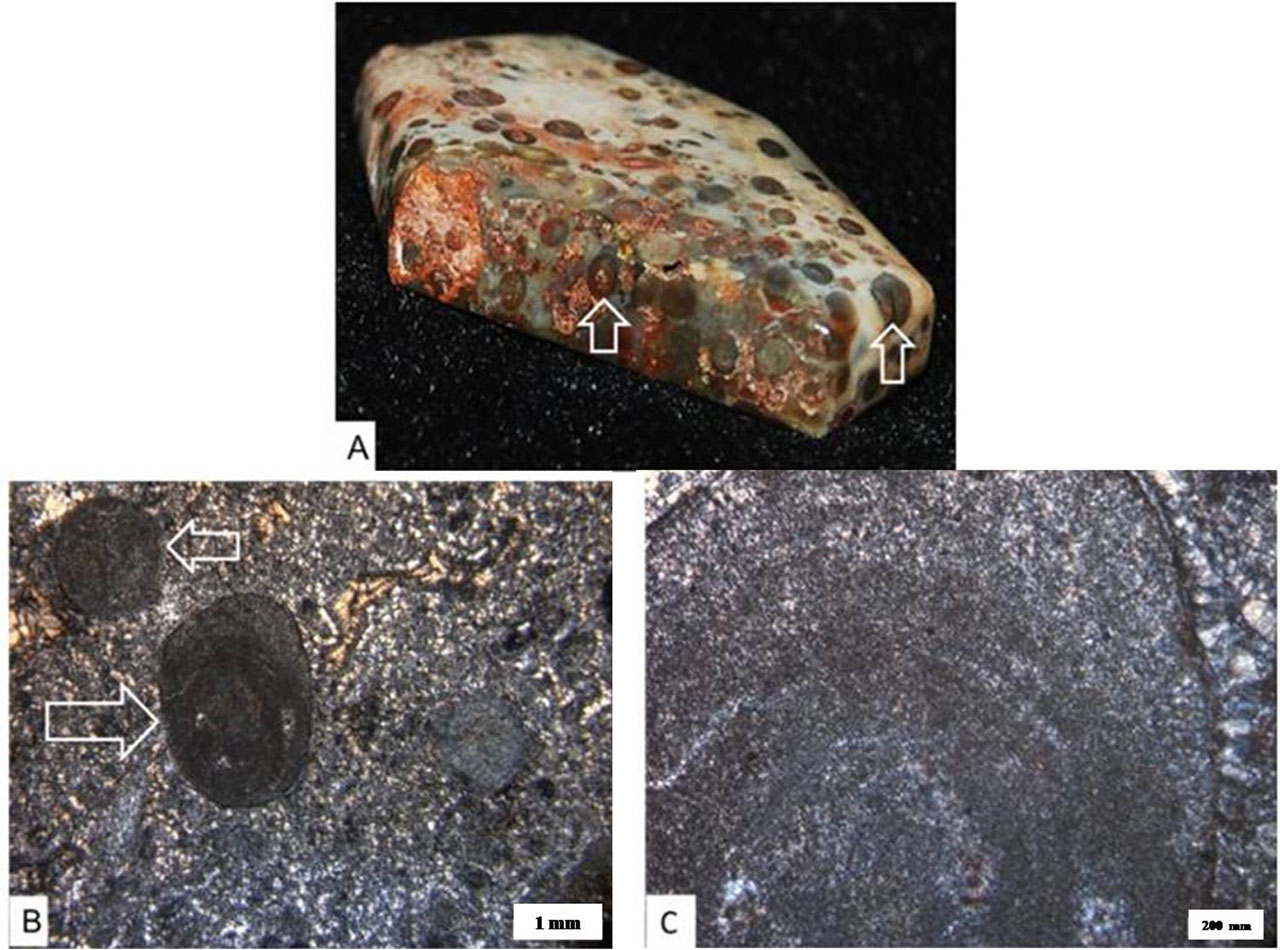
Figure 6. Characteristic textures of Graça agate. (A) Hand-sample, showing a slight banding. The white arrows show the moss texture, both macroscopically and in thin section. (B) Banding and moss texture under the microscope, the first is noticeable because of the Fe staining. (C)Detail of figure B, marking the fine concentric repetition of Fe, Mn oxyhydroxides and chalcedony, as well as the thicker border that separates the botryoidal aggregates from the matrix.
Fe oxyhydroxides
The Fe dendrites are rare in hand samples of Graça chalcedony, and under the microscope can be occasionally seen, when they do appear, they mingle with both fine chalcedony crystals and Mn dendrites, whereas the Fe oxyhydroxides are more developed. SEM/EDS analyses points towards and colloform texture points towards the goethite(Figure7).
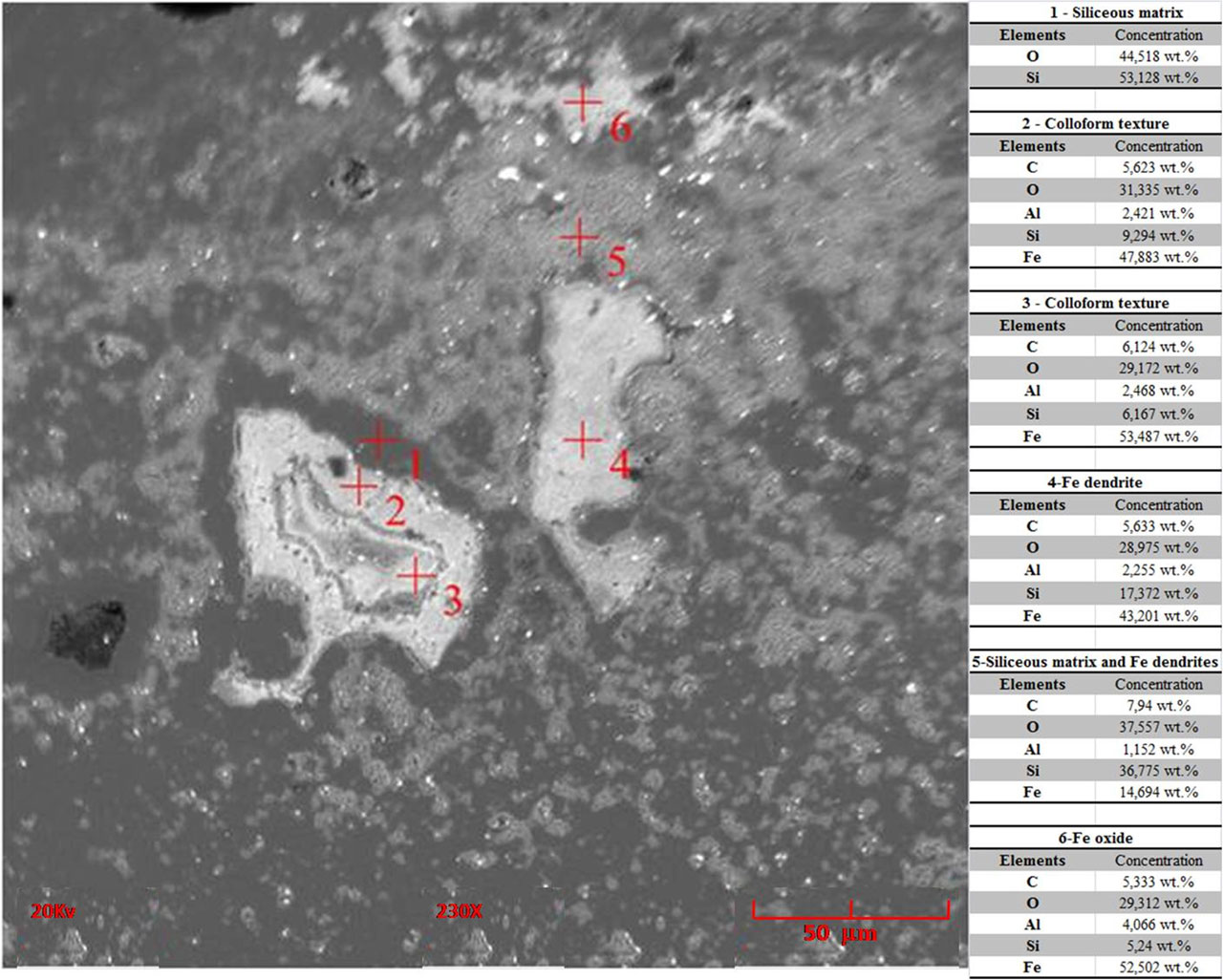
Figure 7: Chemical composition (SEM/EDS) of colloform and massive chalcedony at distinct points on the SEM images. 1. Quartz; 2, 3, 4 and 5. Goethite and quartz with carbon. The colloform texture and dendrites are Always rich on carbon.
Mn oxyhydroxides
These minerals are much more abundant than the Fe oxyhydroxides. Because of their galore they add an exotic appearance to the specimen, and are directly associated to the thinning of the SiO2 matrix, quartz. Some hemi-spherulite aggregates of Mn oxyhydroxides coalesce with both the quartz matrix and the Fe dendrites. SEM-EDS analyses of these Mn are composed of Mn and Ba suggesting to be hollandite (Figure 8), as already identified in the Calcadinha ones by Costa et al (2016).
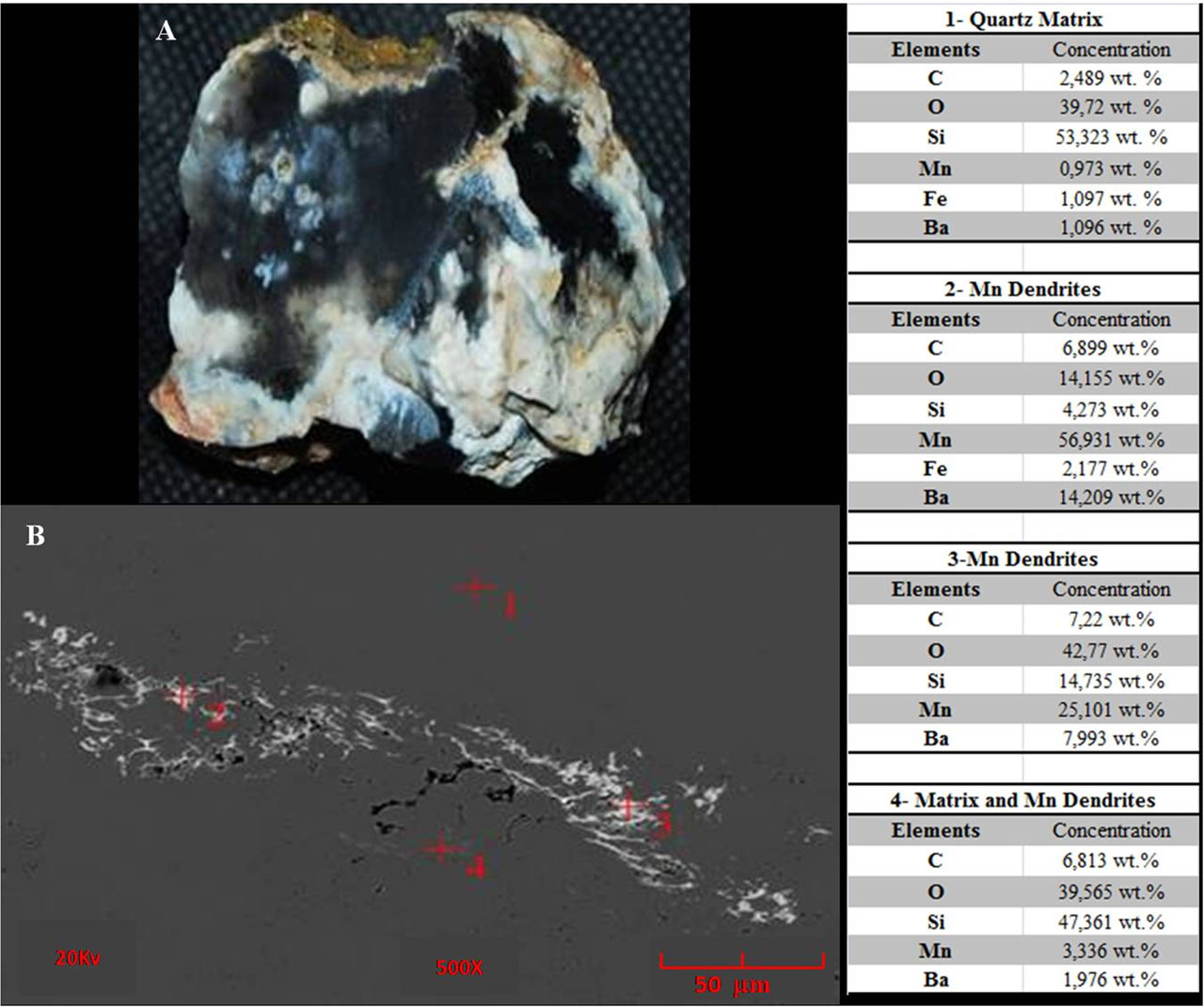
Figure 8. A. hand sample image from where a small fragment was taken for Scanning Electron Microscopy image (B) and EDS semiquantitative analyses. Cryptocrystalline chalcedony matrix (analyzes 1 and 4), Mn oxyhydroxides dendrites domains (analyzes 2 and 3), probably hollandite. The constant presence of carbon is evident at a time in significant values.
Opal
This mineral occasionally appears on hand samples, mingling with remains of sandstone, chalcedony or Mn oxyhydroxides, or filling cavities (Figures 3A and 3B).
Quartz Crystals
Quartz crystals come to light only under sub centimeter size, commonly as druses or overgrown on botryoidal wall and fracture of chalcedonic quartz. Quartz, is also the main component of the relict sandstone, found as part of such chalcedony, therefore may be inherited (Figure 7), similarly to Calçadinha ones (Costa et al., 2016).
.
Microraman analysis
The Raman analysis complements the evaluations and analyzes carried out throughout this article. Three peaks of wavelengths were observed (Figure 9), framed at 220, 430 and 450 cm-1, corroborating with the chalcedony peaks of the program database, the 3 apices occur due to the presence of more than one mineral covered by the laser, the peak of 220 cm-1 corresponds to moganite, the 430 cm-1 to chalcedony and 450 cm-1, quartz (Kingma, 1994). The amplitude of the wavelength indicates that, although the mineralogy is the same, the Chalcedony Graça presents more impurities, irregularities and opacity than the sample inserted in the database of the program.
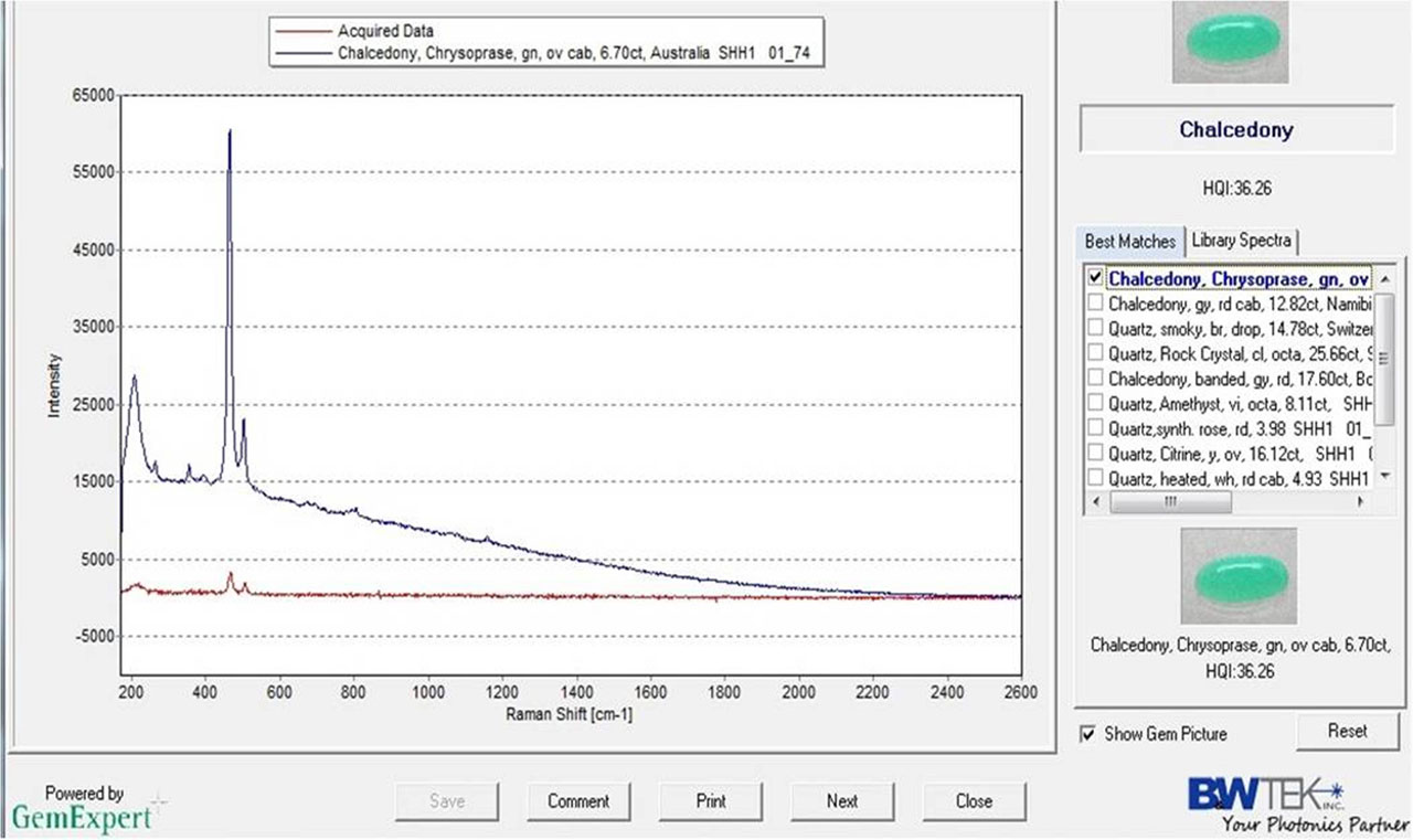
Figure 9- Raman spectrum (red one) for Graça chalcedony sample compared to standard one in database (in blue).
Some gemological aspect of whole chalcedony
Graça chalcedony shows some gemological attributes, such color richness, little banding, sandstone relicts, opal and Fe and Mn oxyhydroxide dendrites (Figure 10). These attributes are influenced by chalcedony internal arrangements, that is, the difference in crystal orientation, some with a-axis parallel to light and some with c-axis parallel causes the light to rapidly diffuse, resulting in some opacity (Jayaraman, 1952).
The relict sandstone, opal, banding, Fe and Mn oxyhydroxides, are striking features, that undoubtedly improve the beauty of Graça chalcedony, seamlessly blending with each other, creating scenarios and images, this is easily exalted by any cut laid upon the gem. Unfortunately, the quasi opacity of Graça chalcedonies limit their gemological use, as complex cuts require that light penetrates into the jewel with as little dispersion as possible (Read, 2005).
Despite its unsuitability towards complex cuts, other types of beautification may still be used, such as dyeing and carving, both are ancient processes that enhance the natural characteristics of chalcedony.
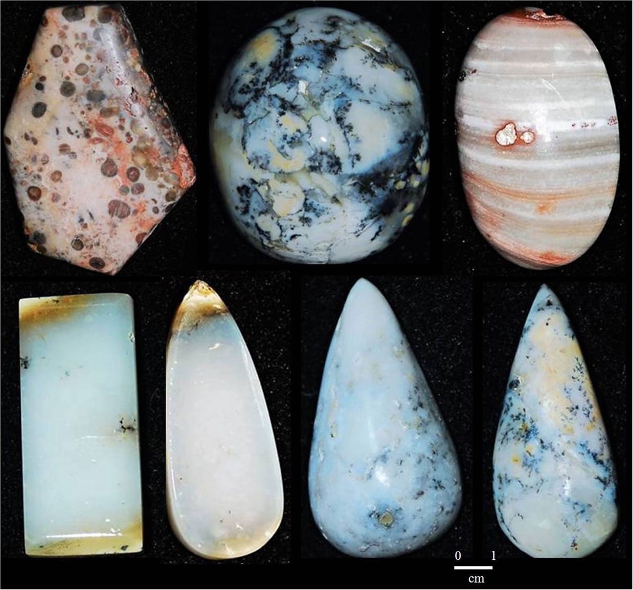
Figure 10. Simple cuts (cabochon, teardrop, rectangular and fantasy) applied to Graça chalcedony, all of them highlight each unique feature in their respective gem.
CONCLUSIONS
Graça chalcedony presents geological occurrence, textural aspects and mineralogical composition, and especially regarding the pervasive presence of Mn and Fe oxyhydroxide dendrite similar to those of Calcadinha, found in Piauí, also in the same geological context. Gemological aspects are also equivalent to each other. They form apparently small occurrences, which so far, as far as we know, do not arouse greater economic interest, but open favorable prospects.
Acknowledgements
The authors acknowledge the Geoscience Institute from Federal University of Pará, specially the Geoscience Museum, for all the support given, analytical or otherwise; LABMEV and the thin section office, also from UFPA; and the São José Liberto museum. Especially to Ms. Graça, the farm owner, who kindly welcomed us and supported the fieldwork. To CNPQ for support scholarship and grant to third author (Nrs.307901/2006-1 and 305015/2016-8), who together with the fourth one carried out the fieldworks. The authors also thank Ubirajara Kimmemgs who kindly improved the illustrations.
REFERENCES
Araújo, R.N. 2015. Depósitos lacustres rasos da Formação Pedra de Fogo, Permiano da Bacia do Parnaíba, Brasil. MS Dissertation, Programa de Pós-Graduação em Geologia e Geoquímica, Instituto de Geociências, UFPA.
Costa M.L., Gomes E.R., Pöllmann H., Angélica R.S. 2013. Opal von Pedro II/Piauí. Aufschluss, 64:49-56.
Costa M.L., Silva Q.A. 2016, Textural patterns, mineralogy, and chemistry of sandstone-related Calçadinha chalcedony (Piauí, Brazil). Brazilian Journal of Geology, 46: 395-409
Kingma, K. J., Russel J. 1994. Raman spectroscopic study of microcrystalline silica. American Mineralogist, 79: 269-273.
Gomes, E.R., Costa, M.L. 1994. Contribuição à gênese da opala de Pedro II – Piauí. Geochimica Brasilensis, 8(1): 79-98.
Gomes, E.R. Costa, M.L. 2007. Opalas do Piauí: Pedro II e Buriti dos Montes (parte II): Diamond New, 8: 54-59.
Marques G.T., Costa M.L., Gomes E.R. 2015. Orange opals from Buriti dos Montes, Piauí: solid inclusions as genetic guides. Rem: Revista da Escola de Minas, 68(1):53-59.
Martins, R. A.; Costa, M. L.; Moraes, M. S. 2010. Floresta fossilizada do Tocantins – uma flora preservada por milhões de anos. 1. ed. NATAL: IFRN EDITORA, 2010. v. 1. 120p.
Morrison, G W., Jaireth, S., Guoyi, D. 1995. Textural zoning in epithermal quartz veins. Townsville, Qld: Klondike Exploration Services.
Moxon, T. & Reed, S.J.B. 2006. Agate and chalcedony from igneous and sedimentary hosts aged from 13 to 3480 Ma: a cathodoluminescence study. Mineralogical Magazine, 70 (5): 485-498.
Moxon, T. 2002. Agate: a study of ageing. Eur. J. Mineral.14, 1109–1118
Moxon, T., Ríos, S. 2004. Moganite and water content as a function of age in agate: an XRD and thermogravimetric study. Eur. J. Mineral. 16, 269–278.
Moxon, T. 2009. Studies on Agate: Microscopy, Spectroscopy, Growth, High Temperature and Possible Origin. Terra Publications, Doncaster, UK, 96 p.
Raman, C.V. & Jayaraman, A. 1955. On the optical behaviour of crypto-crystalline quartz. Proc. Indian Acad. Sci. (1955) 41: 1. https://doi.org/10.1007/BF03050587
Read, P.G. 2005. Gemmology, Third Edition. Published by Elsevier Butterworth-Heinemann.3rd edition.
Santos, M. E. C. M.; Carvalho, M. S. S. C. 2009. Paleontologia das Bacias do Parnaíba, Grajaú e São Luís. CPRM, Serviço Geológico do Brasil. Rio de Janeiro 2009, p.10-18.
Vaz, P. T.; Rezende, N. G. A. M.; Wanderley, J. R. F. & Travassos, W. A. S. 2007. Bacia do Parnaíba. Boletim de Geociências da Petrobrás, 15(2).
Vilasbôas, F. S., Santos, C.R. and Schneider, I.A.H. 2017. Environmental Issues on the Industrial Processing of Raw Agate. Geomaterials, 7, 13-24. http://dx.doi.org/10.4236/gm.2017.71002.